
Simultaneous staining with fluorescently-labelled Annexin V and viability dyes such as 7-AAD or PI can be used to differentiate between early- and late-stage apoptotic events. Annexin V (a Ca++– dependent phospholipid binding protein) binds to phosphatidylserine (PS) on the extracellular membrane. In healthy cells, PS is localized to the cytosolic side of the plasma membrane, but is translocated to the extracellular side in the early stages of apoptosis.
Viability dyes such as 7-AAD and PI rely on loss of membrane integrity for entry into the cell and will not stain cells during early apoptotic events, when membranes are still in tact. As such, cells in the early stage of apoptosis can be identified as positive for Annexin V staining and negative for PI/7-AAD staining, whereas cells in the later stages of apoptosis will be characterized by annexin-V staining in addition to staining with 7-AAD or PI.
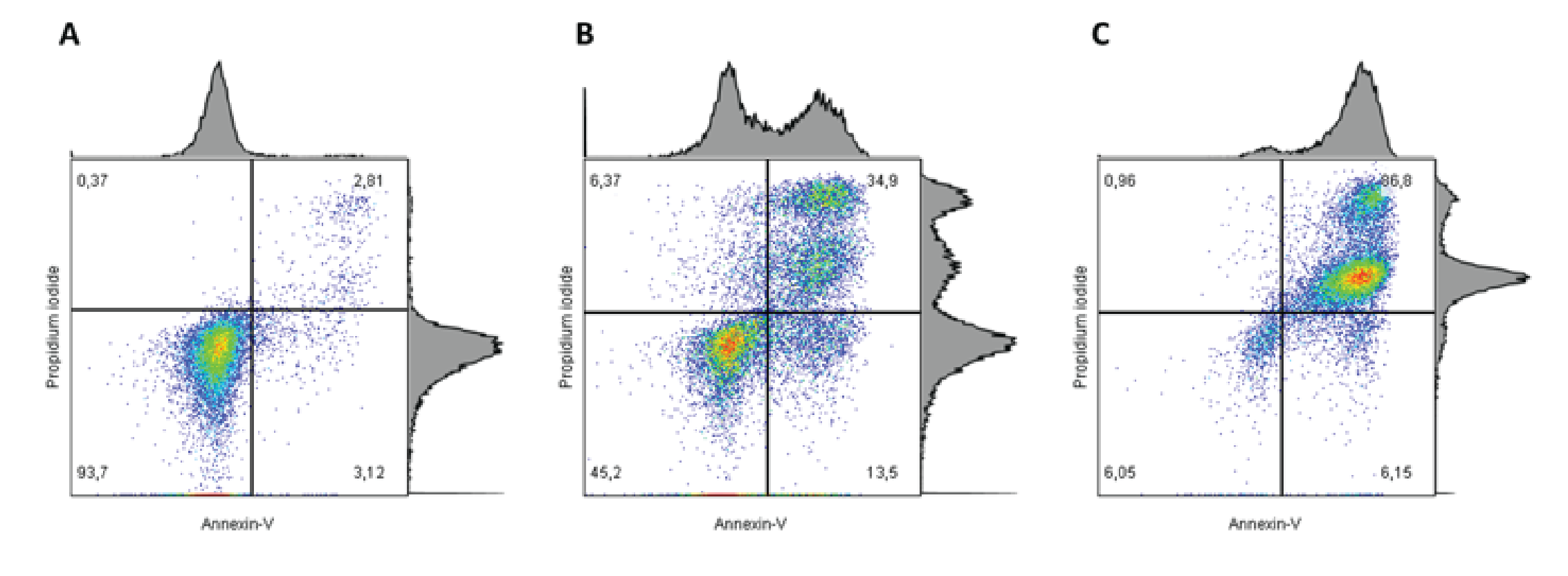
Figure 1: Cells were stained with APC-conjugated Annexin-V and propidium iodide. The proportion of apoptotic events increases with the concentration of drug treatment i.e. from A→C. In addition, a higher proportion of early stage apoptosis is observed with an intermediate concentration of drug treatment (B) relative to the highest concentration (C). The most late-stage apoptotic events are observed with the highest concentration of drug treatment (C).
REFERENCES:
- Ranganathan P, Jayakumar C, Navankasattusas S, Li DY, Kim I, Ramesh G. UNC5B Receptor Deletion Exacerbates Tissue Injury in Response to AKI.Journal of the American Society of Nephrology : JASN. 2014;25(2):239-249. doi:10.1681/ASN.2013040418.
- Yu J, Zheng J, Zhao X, et al. Aminoglycoside Stress Together with the 12S rRNA 1494C>T Mutation Leads to Mitophagy. Lightowlers R, ed. PLoS ONE. 2014;9(12):e114650. doi:10.1371/journal.pone.0114650.
- Yuehong Wang, Jinjun Zhao, Wei Yang, Yayan Bi, Jing Chi, Juanjuan Tian, Weimin Li, High-dose alcohol induces reactive oxygen species-mediated apoptosis via PKC-β/p66Shc in mouse primary cardiomyocytes, Biochemical and Biophysical Research Communications, Volume 456, Issue 2, 9 January 2015, Pages 656-661, ISSN 0006-291X, http://dx.doi.org/10.1016/j.bbrc.2014.12.012.
The loss of mitochondrial function is major apoptotic event characterized by loss of the potential charge across the membrane and can be assessed using JC-1 (a cationic carbocyanine dye). In healthy cells, JC-1 will aggregate within mitochondria and these aggregates will emit red fluorescence at approximately 590 nm. When mitochondria depolarize JC-1 aggregation diminishes resulting in monomeric JC-1 accumulation in the cytoplasm and green fluorescence at 529 nm. This metachromatic shift from red to green fluorescence is easily measured by flow cytometry with blue laser excitation at 488 nm.
REFERENCES:
- Proost I et al. (2008) Functional live cell imaging of the pulmonary neuroepithelial body microenvironment. Am J Respir Cell Mol Biol 39:180-189.
- Liu T et al. (2007) Flex-Hets differentially induce apoptosis in cancer over normal cells by directly targeting mitochondria.Mol Cancer Ther6:1814-22.
- St. John JC et al. (2006) The analysis of mitochondria and mitochondrial DNA in human embryonic stem cells.Methods Mol Biol331:347–74
Another hallmark of apoptosis is the fragmentation of genomic DNA. To monitor DNA breakdown, the 3’-hydroxyl termini of the resultant DNA fragments may be labelled with fluorescent molecule-conjugated deoxyuridine triphosphate nucleotides (dUTPs). The enzyme terminal deoxynucleotidyl transferase (TdT) is added to the assay to catalyze the addition of these dUTPs to the 3’ hydroxyl ends of the DNA fragments. During the analysis an increase in DNA fragmentation will result in the detection of increased fluorescent signal. This assay is referred to as the terminal deoxynucleotidyl transferase dUTP nick end labelling assay or TUNEL assay.
REFERENCE:
Darzynkiewicz Z, Galkowski D, Zhao H. Analysis of apoptosis by cytometry using TUNEL assay. Methods (San Diego, Calif). 2008;44(3):250-254. doi:10.1016/j.ymeth.2007.11.008.
The cleavage of PARP (poly ADP ribose polymerase), a ubiquitous DNA repair enzyme, is another important characteristic of apoptosis. PARP is cleaved by caspases-3,-6 and -7 from its 116 kDa form to two fragments measuring 85 kDa and 25 kDa. The cleaved form of PARP is identifiable through flow cytometry via use of fluorescently-tagged antibodies specifically directed against the cleaved fragments of PARP.
REFERENCE:
Donald D. W. Nicholson et al. Identification and inhibition of the ICE/CED-3 protease necessary for mammalian apoptosis Nature 376, 37 – 43 (06 July 2002); doi:10.1038/376037a0
Caspases are a family of cysteine-aspartic proteases involved in the execution of apoptosis. Caspase activation may be assessed using flow cytometry via various methods including the use of: 1) indirect measures e.g. DEVD peptides and identification of cleaved PARP, 2) FLICA (fluorescent inhibitor of caspases) methodology. The amino-acid sequence DEVD (Aso-Gly-Val-Asp) corresponds to a sequence within the PARP enzyme cleaved by caspase-3, -6 and -7. DEVD sequences conjugated to specific molecules (DEVD-CHO) may be used to inhibit caspase activity and used as an indicators of caspase activity when cleaved. In another example, when DEVD is bound to a nucleic acid binding dye the cleavage of this conjugate releases the the nucleic acid binding dye and the subsequent fluorescent DNA signal is measured using flow cytometry. FLICA reagents include a caspase inhibitor which covalently and irreversibly binds to the reactive cysteines of active caspases. This caspase inhibitor is linked to a fluorescent dye and making it possible to identify the relative extent of caspase activation via the intensity of the fluorescent signal detected by flow cytometry.
REFERENCES:
- Seamus J. Martin, Getting the measure of apoptosis, Methods Volume 44, Issue 3, March 2008, Pages 197–199, Apoptosis doi:10.1016/j.ymeth.2008.02.001
- Scott H. Kaufmann et al., Apoptosis-associated caspase activation assays Methods Volume 44, Issue 3, March 2008, Pages 262-272, Apoptosis, doi:10.1016/j.ymeth.2008.02.001
- Camilla Köhler, Sten Orrenius, Boris Zhivotovsky, Evaluation of caspase activity in apoptotic cells, Journal of Immunological Methods, Volume 265, Issue 1-2, July 2002, Pages 97- 110, Evaluation of Apoptosis. doi:10.1016/S0022-1759(02)00073-X
Please see the following resources and references for more application details.
- eBioscience: Apoptosis Research Product Guide
- BD Biosciences Apoptosis Applications Brochure
- Fishser Scientific – Molecular Probes Apoptosis / caspase Assays for Flow Cytometry
- Denovo Software – Flow Cytometry Text Book
- Wlodkowic D, Skommer J, Darzynkiewicz Z. Flow cytometry-based apoptosis detection. Methods in molecular biology (Clifton, NJ). 2009;559:10.1007/978-1-60327-017-5_2. doi:10.1007/978-1-60327-017-5_2.
- Wlodkowic D, Telford W, Skommer J, Darzynkiewicz Z. Apoptosis and Beyond: Cytometry in Studies of Programmed Cell Death. Methods in Cell Biology. 2011;103:55-98. doi:10.1016/B978-0-12-385493-3.00004-8.